

Larval Size Photographs Protocol
This protocol describes the process for taking photographs of preserved coral larvae for measurement of larval size (volume, surface area, length, etc.).
Protocols developed by Danielle Becker and modified by Ariana Huffmyer.
Materials Required
- Pipette
- Microscope dish
- Scale bar
- OMAX compound light microscope
- OMAX microscope camera
- OMAX camera software
- Samples preserved in either Z-Fix or 4% paraformaldehyde
Procedure
- Remove an aliquot of preserved sample from tube after gentle mixing by pipette. CRITICAL: Only use equipment that is meant for use with fixatives. If new equipment is used, dispose of properly as hazardous waste or label as “fixative only” for future use.
- Place an aliquot under the microscope in the microscope dish such that 20-30 individuals are in view. If the photograph frame is too small for visualization of 20-30 samples, reduce aliquot size and take multiple photographs per tube. Multiple photos can be taken per sample.
- Ensure that the scale bar is included in the photograph.
- Record photograph number on datasheet along with tube number and replicate number (if multiple samples are taken). Rename photographs with sample name and replicate number. For example (Sample#-Photo1, Sample#-Photo2, etc.).
- Upload photographs to Google Drive
- Repeat for each sample
- Samples can then be analyzed for size or volume in ImageJ/Fiji software.
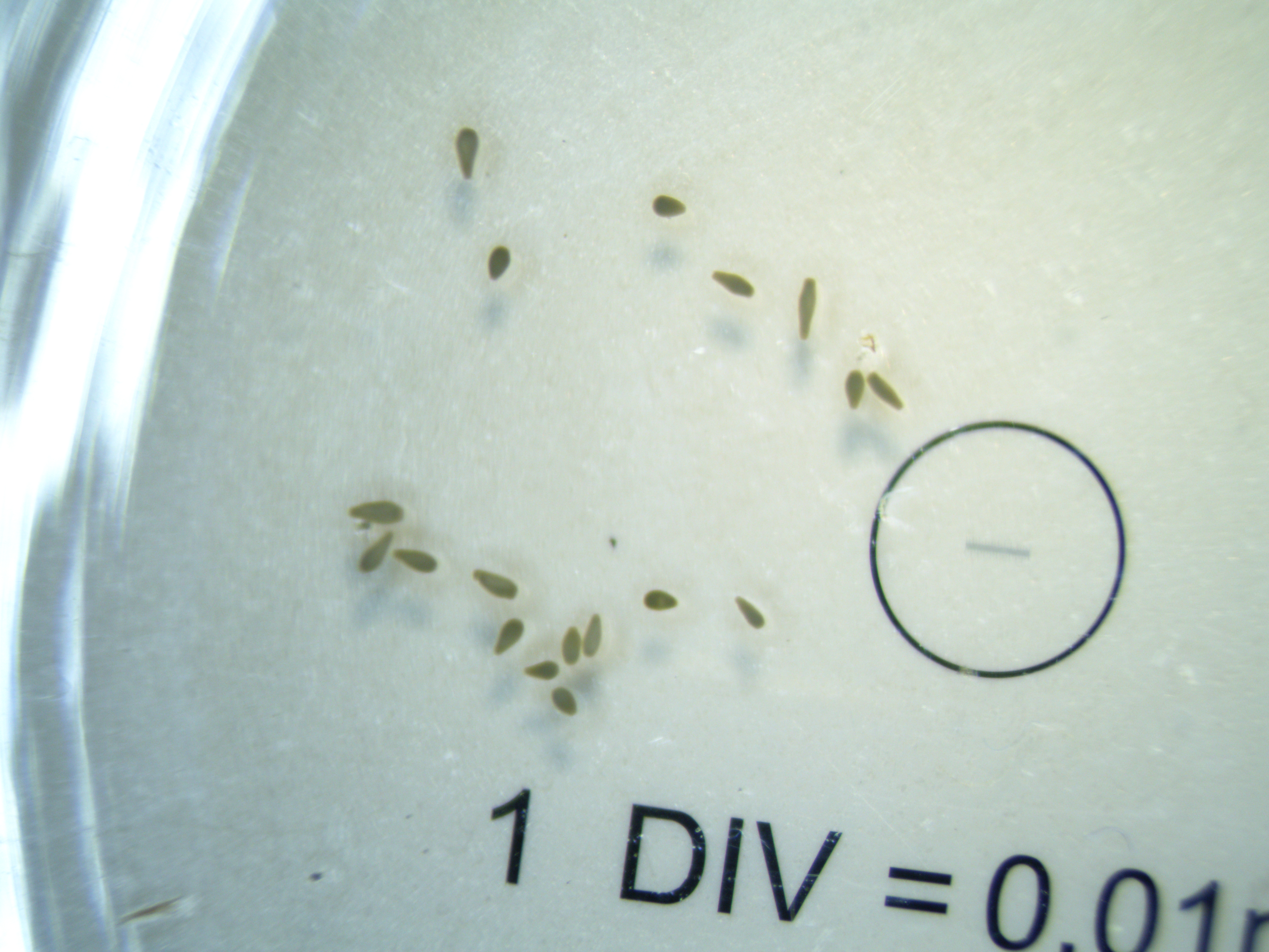
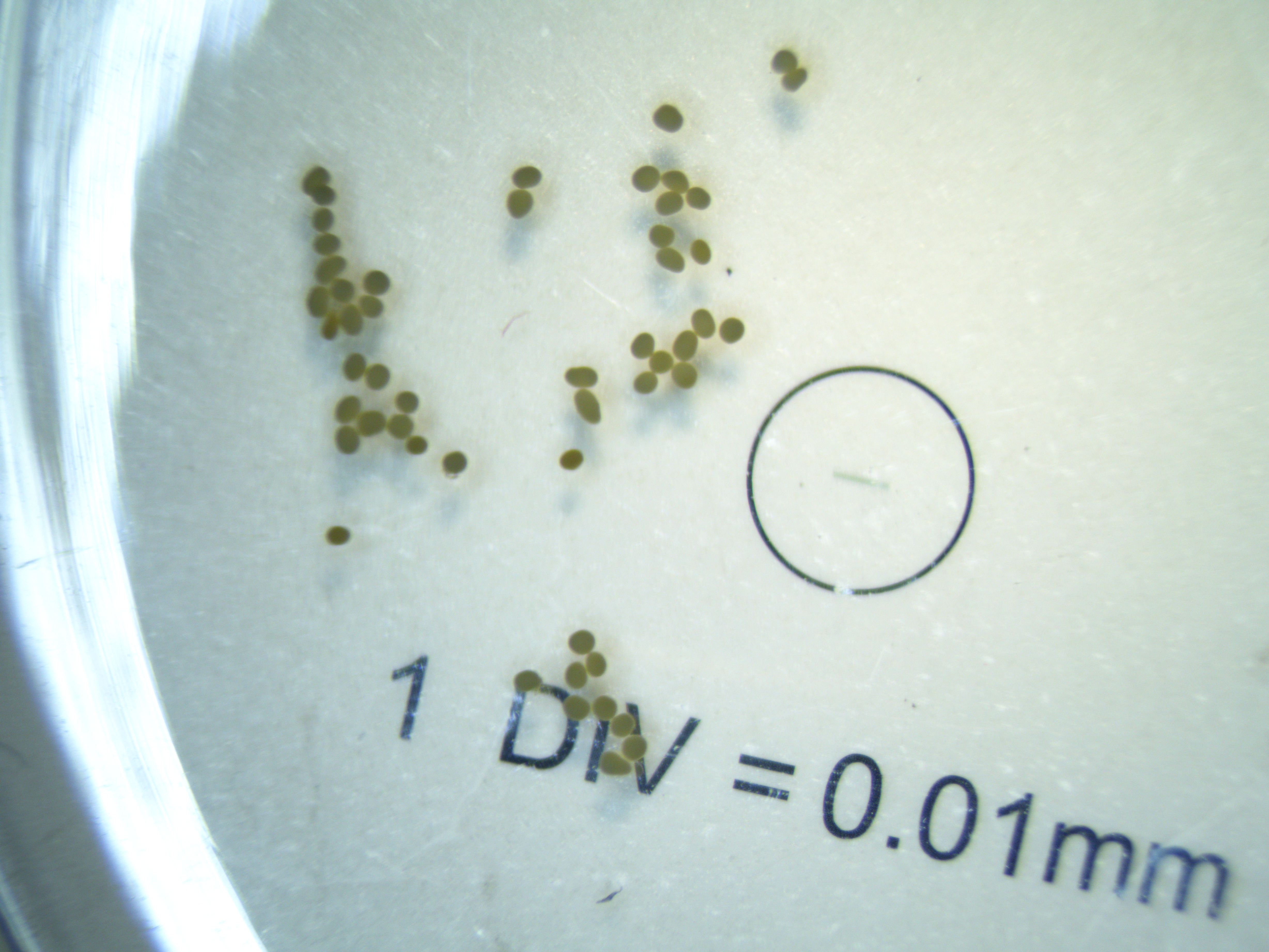
See an example photograph below.
Measurement in ImageJ
- Open ImageJ or Fiji software
- Open the first photograph
- Set scale for each picture individually using the stage micrometer in the picture (1mm). Write down the units used for the scale (mm, cm, etc). In our photographs, the scale line = 1.0mm. The small divisions (n=10) within the larger line are 0.1mm.
- We will measure size for n=20 larvae per photograph (if there are less than 20 larvae, measure all that are in the picture).
- Use command + to zoom in on the image and use command - to zoom out. Zoom in to an individual larvae.
- For each larvae, use the line tool to draw a line on the larvae. Then click command+m. This will measure the longest length. Record “Length” in data sheet from the report that shows in ImageJ/Fiji. The longest length should equal approximately 0.400-0.500 mm.
- Then, on that same larvae, measure the perpendicular width to the length you just measured. Record “Length” on data sheet.
- Larval volume will then automatically calculate in the datasheet.
- Repeat for all larvae (n=20 per photo).
- On data sheet, record necessary metadata such as photograph ID, date, and any notes.
Written on April 13, 2022